RadOnc News

FLASH-RT and the Mechanics of Ultra High Dose Radiotherapy

FLASH-RT, a glimpse into the future of high dose rate radiation oncology. What is it, where it is now, and what lies in store?


Treating cancer has long bound the radiation oncology community in a struggle between maximizing dose to a target volume while simultaneously sparing healthy tissues. Until recently, this was achieved using a low dose rate over many fractions. In doing so, tumors reoxygenate, and healthy tissue recovers, reducing short- and long-term acute effects.

Of course, studies have indicated that cancers with low α/β ratios respond better to high dose (2+ Gy/Fraction) hypofractionated treatments. Since the delivered dose is critical in tumor sterilization, many contemporary techniques such as VMAT, adaptive radiotherapy, and hypofractionation seek to increase the fractional dose to the target while also striving for a tighter margin. However, these delivery techniques lead to a low dose bath to surrounding healthy tissue volumes. One promising new solution to this phenomenon is FLASH radiotherapy.


Brief History

FLASH is a delivery technique that uses ultra-high dose rates of 40+ Gy/s, orders of magnitude higher than conventional dose rates. Initially described in 1966 by Hornsey and Alper, the FLASH effect reduces radiation-induced toxicities and spares healthy tissues while maintaining similar tumor response as conventional rates.

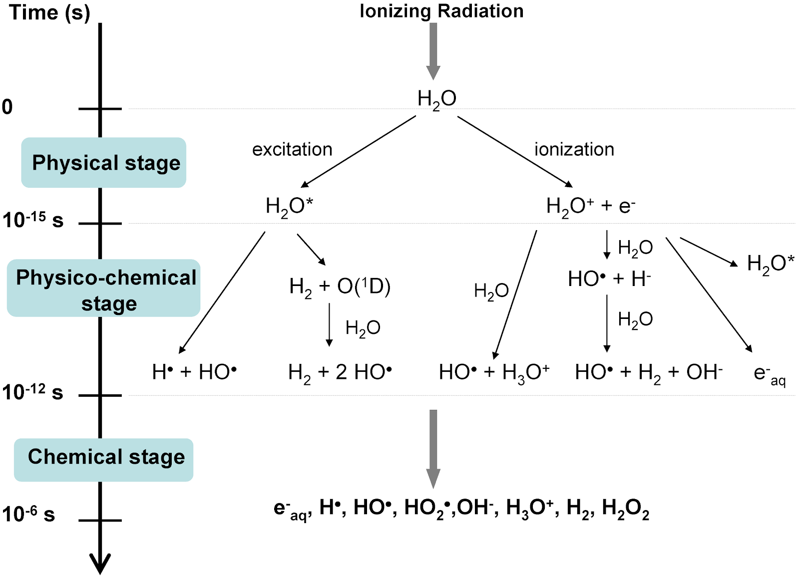
Hornsey and Alper attributed the viability of cells irradiated with ultra-high dose rates to a state of induced transient hypoxia, which minimized the yield of reactive oxygen species (ROS). Hypoxic states protect against low-LET radiation since oxygen acts as a radiosensitizer. ROS pose a problem in healthy tissues as they damage DNA and RNA and can oxidize amino acids and enzymes, leading to a breakdown in cellular function. Unfortunately, their work was left mostly unstudied since many thought the doses needed to induce hypoxia locally were too high.

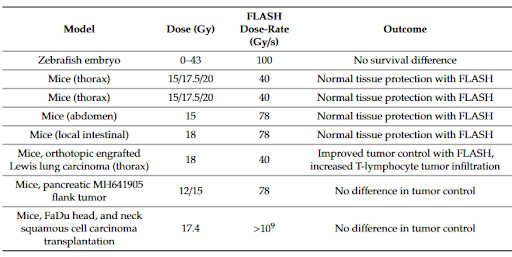
Favaudon et al. reversed this belief in 2014 when they described a similar dose rate effect on physiological response in mice treated using conventional dose rates (0.3 Gy/s) and ultra-high dose rates (40 Gy/s). Their results indicated that FLASH showed similar repression of tumor growth while also significantly reducing the occurrence and severity of early and late-stage acute toxicity. Unlike Hornsey and Alper, Favaudon et al. did not attribute the effects to a state of transient hypoxia. Instead, they suggested a change in DNA repair signaling might play a role.

Their findings sparked a resurgence of research on the subject to unravel the mysterious mechanism(s) behind these effects. To date, despite 3000 papers having been published on the topic, the community does not appear to have arrived at a consensus. According to Dr. Emil Schüler, who co-leads the FLASH initiative at MD Anderson, “While the oxygen hypothesis gets a lot of attention, it cannot be uniquely responsible for the effects we see since oxygenated tumors undergoing FLASH are still responding similarly to conventional delivery. It is likely due to an interplay of oxygen depletion, reactive oxygen species and lymphocyte sparing.”


Deep Dive into Mechanisms

When incident ionizing radiation interacts with biological material, it deposits its energy through direct and indirect events. Direct interactions account for 33% of cell damage and involve direct ionization of molecular structures (primarily DNA). Indirect interactions make up the remaining 67% of damage and follow a series of chain reactions to produce ROS, free radicals, and aqueous electrons from water as shown below. The ROS and free radicals can cause a variety of complex lesions on DNA to form and create damaging organic radicals.


Oxygen Effect

In conventional radiotherapy, the tissue is given a chance to repair between fractions, allowing tumor cells to reoxygenate, which perpetuates their radiosensitivity. Conversely, FLASH pulses occur faster than the re-oxygenation kinetics process, and the entire treatment is delivered in a single fraction. This high dose and lack of re-oxygenation might be the key to the dose sparing effects seen. Since the number of ion pairs formed is proportional to the dose rate, a single 4-6MeV/MV pulse of 10Gy FLASH creates around five orders of magnitude more ionization events than a conventional 6MeV electron or 6MV photon beam pulse. Ionization events cause local oxygen consumption, and the high dose rate is thought to induce a state of transient hypoxia and radioresistance in irradiated tissue. Assorted publications have correlated oxygen concentration and tissue sparing by demonstrating that highly oxygenated cells required an increased dose to exhibit the FLASH effect.
 

Reactive Oxygen Species

While only a handful of studies have covered FLASH’s efficacy in tumor control, similar efficiency for tumor repression has been demonstrated. Figure 1 summarizes the results of a few studies using proton FLASH. Researchers believe that a differential biochemical state between FLASH and conventional RT is at play to maintain a similar tumor control while inducing radioresistance. Spitz et al. hypothesized that Fenton-type reactions could account for the differential response in ROS damage. Fenton reactions occur when hydrogen peroxide (H 2O 2) oxidizes Iron (Fe³⁺ or Fe²⁺), generating additional free radicals that destroy organic compounds and creating a cascade of additional peroxidation chain reactions. In tumor cells, they found the concentration of labile iron (protein-bound iron, not free) and transferrin receptor activity to be 2 to 4-fold higher than normal cells. Also, normal tissues have a larger ability to control labile iron and remove the FLASH induced hyperoxides.
 

Immune Response

The final mechanism of note is the immune system response to radiation. As immunotherapy has shown, lymphocytes play an essential role in tumor control. However, lymphocytes are susceptible to chromosomal aberrations when irradiated. Yovino et al. found in a conventional delivery of an 8cm GBM treated with 200cGy x 30 that 99% of the blood pool was exposed to 0.5 Gy or more, a potentially lymphotoxic dose. FLASH delivery would reduce the pool of blood exposed. Furthermore, Rama et al. found increased CD3+ T lymphocyte recruitment from tumor periphery to tumor core in mice treated with FLASH compared to conventional treatment.
 

FLASH in the Clinic?

What would FLASH look like in the clinic? While we are far from clinical use, Dr. Schüler hypothesizes that high energy electrons are the most probable solution. Many electron modes already have enough current for FLASH dose. High energy electrons (100-200 MeV) would allow for treatment of deep tissue lesions without the need for photons. Because normal tissue is largely spared with FLASH, the treatment would likely not require much dose shaping with MLCs or rotational techniques, and minimal intrafraction motion management is necessary.

Another topic of clinical interest is dosimetry. With such high doses, ion chambers saturate and diodes fail, leaving only film, TLDs, and other dose rate independent methods for measurement. To deliver a specific dose, film is used to determine the dose per pulse and the desired number of pulses must be calculated since MU chambers saturate. As we look towards the future and clinical implementation, QA and dosimetry will be important areas of investigation.


Conclusion

In recent studies, FLASH appears to offer advantages in dose escalation, tissue sparing, and the treatment of radioresistant tumors. Numerous animal studies have shown effects like reduced fibrosis incidence in lung treatment, reduced neural structure degradation, and limited skin toxicity. However, it is far from a clinical application, and the underlying mechanism for the results remains a mystery. At present, current research is focused on toxicity for phase I trials and potentially dose escalation. Depending on study and trial outcomes, we can begin to look forward towards clinical adoption.


Leave a comment